by Vickie Byard, CVT, VTS (Dentistry)
This morning, we performed dentistry on a 10 year old Rat Terrier. I just love showing off our dental surprises.
We performed the cleaning and charting of this mouth. There was a periodontal pocket evident on the distal root of the left lower molar. Here is the clinical picture after the cleaning: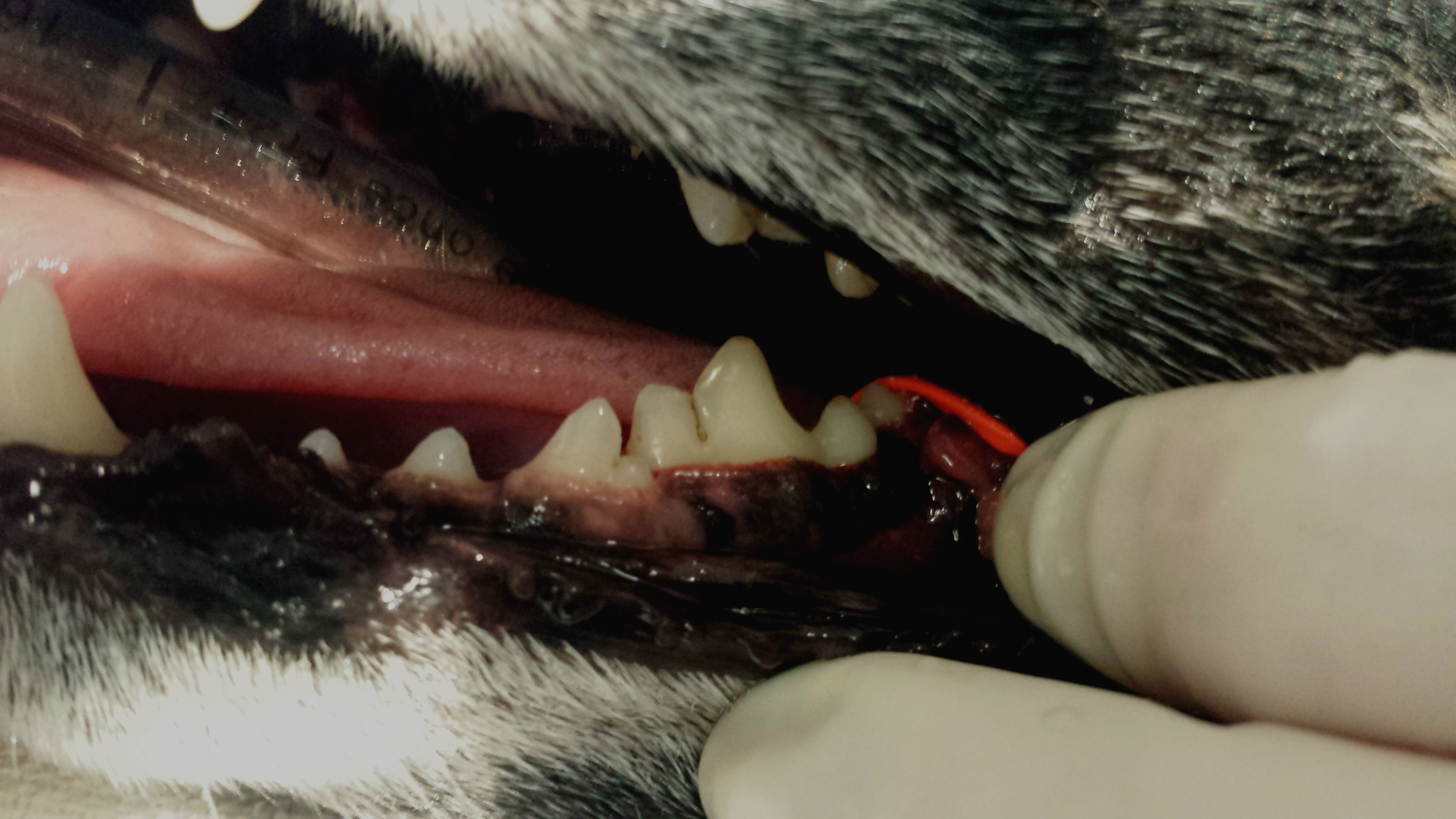
Clinically, there is not much evident or alarming in this image.
The following was the startling radiograph of the same tooth.
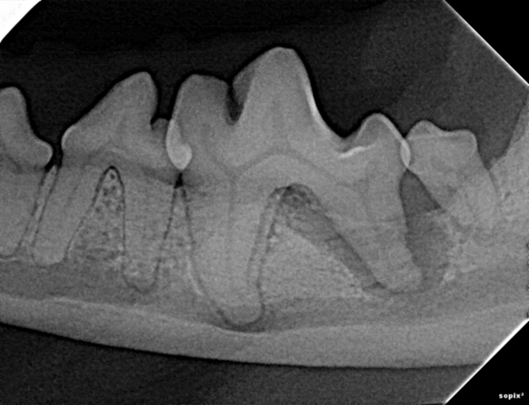
With this new information, the veterinarian was able to make an informed decision as to the best way to proceed. Today was this dog’s lucky day. Despite the fact that initially this owner felt that there was nothing wrong, with proper education they gifted this dog with the extraction of the lower first and second molars. I expect that this pet will be acting like a puppy again within days.
Dr. Marty Becker, Kim Campbell Thornton and Mikkel Becker describe some outward signs of discomfort that pet owners can watch for related to dental problems in an article published just today. They also wisely state that, “Pets don’t have any way of telling us that something is wrong, and it’s natural for them to hide signs of weakness or pain so they don’t become targets of predators.”
If you are working in a small animal practice or own a small animal practice and do not yet have intraoral radiology capability, feel free to contact me if you would like to consider adding such a valuable service. It is hard for me to contain my enthusiasm for the benefits of this single piece of medical equipment.
*If any of this information was useful or you would like to see similar content, “Like” the Pet ED Veterinary Education and Training Resources Facebook page and sign up on our website for our newsletter.